Amniotic fluid: what is it and what is it used for in pregnancy?
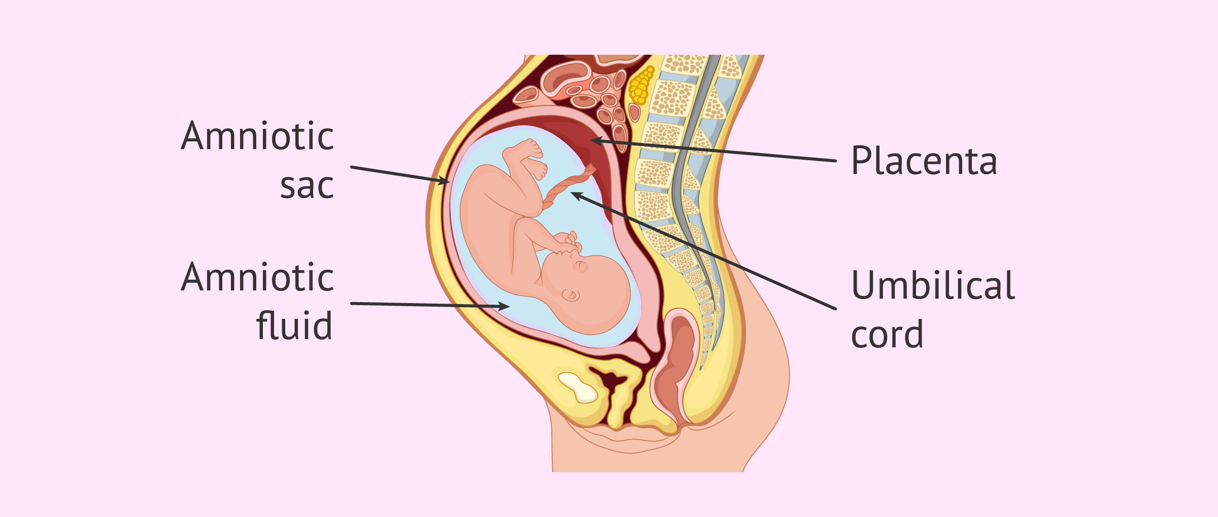
Amniotic fluid is the fluid that surrounds the fetus during pregnancy inside the amniotic sac, the sac where the fetus grows and develops until birth.
During gestation, the amniotic fluid has very important functions, as it is what sustains and protects the fetus.
Any alteration in the amniotic fluid poses a risk to the pregnancy and should therefore be monitored by ultrasound, especially in the last trimester.
The different sections of this article have been assembled into the following table of contents.
Contents
- 1.
- 2.
- 3.
- 3.1.
- 3.2.
- 4.
- 4.1.
- 4.2.
- 4.3.
- 4.4.
- 5.
Composition
Amniotic fluid begins to form around the fourth week of pregnancy when the embryo has already been implanted in the uterus and the amniotic sac, also called the amnion, is formed.
The composition of amniotic fluid varies throughout pregnancy. In the first trimester, amniotic fluid is an ultrafiltrate of maternal blood plasma and is composed of proteins, carbohydrates, carbohydrates, carbohydrates, and electrolytes that will help fetal development.
From week 12 onwards, the fetus is also involved in the renewal of the amniotic fluid by providing its urine, which will be the main component in the coming weeks.
The amniotic fluid is regenerated and is in continuous circulation: the fetus ingests and expels it several times a day.
The amount of amniotic fluid also changes throughout gestation. Around week 14, the fluid volume is about 100 ml.
The maximum amount is reached in the 34th week, where the fluid volume is about 800-1000 ml.
From the 38th week of pregnancy, this amount begins to decrease to approximately 600 ml.

Functions
During pregnancy, the fetus is floating inside the uterus suspended by amniotic fluid. This is essential for the correct development of the fetus and gestation.
The main functions of the amniotic fluid are as follows:
- It allows the fetus to move freely without the membranes of the amniotic sac adhering to its body. This contributes to proper bone growth.
- Protects the fetus from external shocks or sudden movements. The fluid cushions possible abdominal trauma to the mother and/or the effect of uterine contractions.
- In the same way, it also cushions the movements of the fetus so that the mother does not feel pain. In addition, the fluid prevents possible damage to nearby maternal organs, as well as compression of the umbilical cord.
- It maintains an adequate and constant temperature around the fetus, preventing heat loss, in addition to providing the most suitable sterile environment for its development.
- It allows for the proper development of the fetal lungs.
- Finally, it helps to accommodate the fetus to the birth canal when the water has not yet ruptured and, when the water breaks, the amniotic fluid lubricates the birth canal.
It is a very important fluid during fetal development, without which pregnancy could not take its course.
Amniotic fluid assessment
Assessment of the amount of amniotic fluid during pregnancy is an indicator of fetal well-being. It can be done by ultrasound sonography, although it requires the gynecologist to be highly experienced.
Assisted procreation, as any other medical treatment, requires that you rely on the professionalism of the doctors and staff of the clinic you choose. Obviously, each clinic is different. Get now your Fertility Report, which will select several clinics for you out of the pool of clinics that meet our strict quality criteria. Moreover, it will offer you a comparison between the fees and conditions each clinic offers in order for you to make a well informed choice.
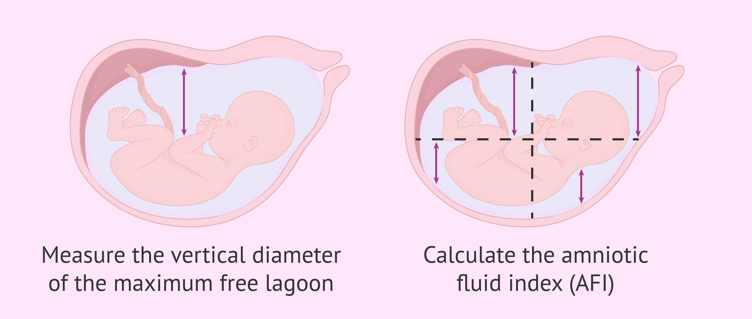
There are two methods used for the estimation of amniotic fluid volume:
- Measure the vertical diameter of the maximum free lagoon
- consists of making a single measurement of the largest quadrant of amniotic fluid that is free of fetal parts and umbilical cord. The normal size of this diameter is between 2-8 cm.
- Calculate the amniotic fluid index (AFI)
- consists of dividing the uterine cavity into four quadrants and then measuring the diameters of the maximum free amniotic fluid gaps in each quadrant. The sum of these four measurements will give the AFI, whose normal value is between 8-24 cm. This technique is also known as Phelan's method.
Abnormal amount of amniotic fluid
Amniotic fluid volume is measured in the third trimester of pregnancy. A value outside the ranges established as normal indicates that there is some alteration in the amount of amniotic fluid.
Too little amniotic fluid, as well as too much volume within the amnion, can cause problems for both mother and fetus. These alterations are as follows:
- Oligohydramnios
- is the presence of low amniotic fluid due to an AFI<8. It may be due to genitourinary anomalies or fetal malformations. Maternal hypertension or utero-placental insufficiency are also causes of oligohydramia.
- Polyhydramnios
- refers to excess fluid in the amniotic sac due to an AFI>24. It may be due to multiple pregnancy, congenital anomalies, gestational diabetes or maternal infections.
Generally, pregnancies in which one of these anomalies is detected, develop normally, result in a normal delivery and the birth of a healthy baby.
However, there are exceptional cases in which these alterations in the amniotic fluid may pose a risk. Therefore, it is necessary to carry out additional and specific monitoring of the pregnancy.
For more information on this, we recommend you read the following article: Amniotic fluid alterations.
Amniocentesis
Amniocentesis is a prenatal test that can be performed between 14 and 20 weeks of pregnancy to evaluate the health status of the fetus.
It consists of extracting a sample of amniotic fluid for analysis, as it contains fetal skin or kidney cells.
The information provided by amniocentesis is as follows:
- Sex of the fetus
- If there is malformation in the neural tube
- Genetic alterations in chromosomes
- Fetal pulmonary maturity status
- Possible hereditary metabolic or muscular diseases
However, amniocentesis is a test that carries a risk for the fetus and, therefore, will only be performed in cases of suspected malformation or chromosomal alteration.
The amniocentesis test is recommended to perform in women over 35 years of age.
For more detailed information on this, you can continue reading here: What is amniocentesis?
FAQs from users
What are the characteristics of amniotic fluid?
Amniotic fluid is a fluid that is light in color, almost transparent and slightly yellow. Gradually, the fluid may take on lumps from the peeling of the fetus's skin. Once past the due date, the amniotic fluid turns milky.
On the other hand, if the liquid darkens or turns green, it means that the fetus has released intestinal content, known as meconium. This poses a risk to the fetus, as the meconium can cause damage to his lungs if he breathes it in.
What is the function of amniotic fluid?
The main functions of amniotic fluid are to protect the fetus and the surrounding maternal organs, as well as to maintain its correct homeostasis and body temperature.
How can I recover amniotic fluid in pregnancy?
This depends on the stage of pregnancy and the cause of the oligohydramnios. In general, it is recommended to maintain plenty of rest, drink plenty of water and isotonic drinks, eat more vegetables and juicy fruits such as melon, orange, or grapes.
What happens to babies born in the amniotic sac?
This is known as veiled delivery. It occurs when the baby is born with the sacs intact and unruptured and the baby is surrounded by amniotic fluid. This happens rarely, usually during the course of a natural delivery.
When the amniotic sac does not rupture on its own during delivery, artificial rupture is performed by the physician.
Recommended readings
Occasionally, amniotic fluid may leak out of the sac due to a rupture or fissure, which is always a cause for concern. You can read more about this topic in the following article: How do I know if I am leaking amniotic fluid in pregnancy?
We make a great effort to provide you with the highest quality information.
🙏 Please share this article if you liked it. 💜💜 You help us continue!