The process of fertilization is well known in society. The sperm fertilizes the egg in the fallopian tube and the embryo then descends into the uterus where it implants in the endometrium.
However, there may be a lack of knowledge about how the extraembryonic structures of the placenta and umbilical cord originate. In addition, some of the gestational complications that often occur are related to these structures.
Provided below is an index with the 7 points we are going to expand on in this article.
- 1.
- 2.
- 2.1.
- 3.
- 4.
- 4.1.
- 4.2.
- 4.3.
- 4.4.
- 5.
- 6.
- 7.
Trophoblast formation and implantation
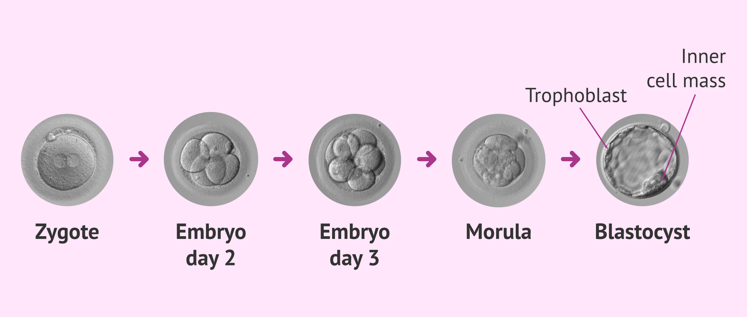
After fertilization, the embryo begins to divide, from one cell to two, from two to four, from four to eight, and so on. Each of the cells of the embryo divides in two.
From day 3 of embryonic development, the blastocyst begins to form, the embryonic structure necessary for implantation in the uterus and, therefore, pregnancy.
For this, the embryo has to change from being compacted to having a structure with an internal cavity and two well-differentiated cell groups. Thus, when the embryo implants in the endometrium, around the 5th or 6th day after fertilization, two layers can be clearly distinguished:
- Trophoblast
- will give rise to the placenta and the umbilical cord. The cells that form the trophoblast, in turn, will differentiate during the second week of pregnancy into 2 layers of cells, an inner one called cytotrophoblast and an outer one called syncytiotrophoblast.
- Inner cell mass
- from here will derive all the tissues that will form the embryo. Specifically, the transformation of the inner cell mass of the blastocyst into an embryonic disc, which is the beginning of all tissues and organs, takes place.
For embryo implantation to take place, the syncytiotrophoblast is responsible for digesting the endometrium by proteolytic enzymes. In this process the blood vessels of the endometrium will be ruptured and rapid invasion by the syncytiotrophoblast will occur.
Chorion and placenta formation
Once embryo implantation is complete, the mother's blood vessels will be fully connected to the syncytiotrophoblast.
This union of vessels between the embryo, the mother and the syncytiotrophoblast will give rise to a structure called the chorion. This structure together with the mucosal wall of the uterus will form the placenta. In this way, a utero-placental circulation will be established thanks to the formation of deep villi, which branch out.

The placenta is a round-shaped organ that usually measures about 22 cm in diameter and is 2.5 cm thick. The weight of the placenta is usually around 500 grams, although this weight does not include the membranes or the cord. In addition, a fetal surface or chorionic plate and a maternal surface or basal plate are distinguished in the placenta.
What is the function of the placenta?
The placenta is responsible for providing oxygen and nutrients to the baby throughout its development and, therefore, throughout the pregnancy. In addition, this structure has an immunological action, since the placenta allows the embryo not to be recognized as foreign.
Another function of the placenta is to synthesize hormones and growth factors. These molecules are essential for the baby's metabolism, but also for the mother. One of the hormones produced by the placenta is chorionic gonadotropin or more popularly known as beta hCG.
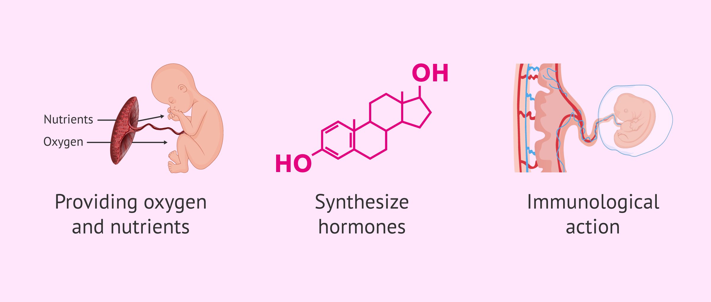
Apart from this hormone, the placenta also secretes estrogens, progesterone, relaxin and other female hormones (GnRH, TRH, inhibin, etc.). The development of placental lactogen, a hormone that promotes fetal growth, lactation and the production of other hormones such as prolactin, also occurs.
How is the umbilical cord formed?
The embryo will grow in turn, although more slowly than the external structures. Embryonic growth depends on the influx of nutrients and oxygen, as well as the elimination of waste products.
To connect the embryo and the trophoblastic envelope, the so-called fixation pedicle is formed, which will later become the umbilical cord.
The umbilical cord is the connection between the placenta and the fetus. Its composition consists of two arteries leaving the fetus towards the placenta and a vein leaving the placenta towards the fetus.
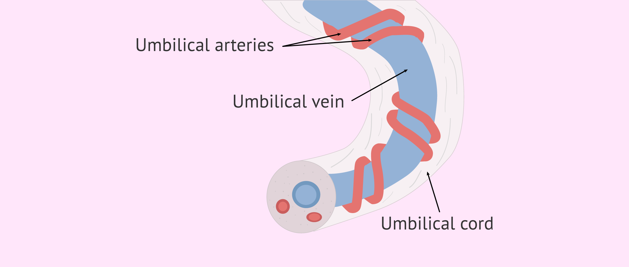
Thus, there is no direct exchange of blood between the mother and the embryo, everything is done through the chorion and the umbilical cord. These structures also serve as a filter, offering protection to the fetus against possible harmful agents present in the maternal blood.
Although the origin of the umbilical cord and placenta is extraembryonic, maternal tissues are needed to form the entire maternal-fetal set. This set will serve during the nine months of pregnancy for the fetus to nourish itself and exchange the necessary gases during embryonic development.
FAQs from users
What is embryo implantation?
Embryo implantation is the process by which the embryo joins the internal uterine wall known as the endometrium. The embryo is able to penetrate the uterus. From this moment on, the formation of the future placenta will begin, which will provide nutrition, sustenance and protection for the pregnancy.
When does placenta formation begin?
The placenta is a structure that is formed at the time of embryo implantation in the uterus. Therefore, the formation of the placenta usually takes place in the week of pregnancy and continues to evolve until the third or fourth month of gestation. At this point, the placenta is fully formed, but it may undergo some changes until the end of the pregnancy.
What hormones does the placenta produce?
The placenta has an endocrine action. One of the main hormones produced by the placenta is human chorionic hormone or hCG.
However, hCG is not the only hormone; the placenta is also capable of producing human placental lactogen (hPI), thyroid hormones and steroid hormones. This hormone production by the placenta takes place around week 12 of pregnancy.
Does the placenta or the umbilical cord form fir
The formation of the placenta and the umbilical cord takes place at the same time. What happens is that the union of the placental capillaries will form the three blood vessels that form part of the umbilical cord; that is, the two arteries and the vei
Suggested for you
One of the possible gestational complications that can occur is placental abruption. If you want to know more about this topic, we recommend you visit the following article: What is placental abruption and why does it occur?
Aside from problems related to the placenta, problems can also occur in the umbilical cord. In this case, you will find more information in the following link: What complications related to the umbilical cord can occur?
We make a great effort to provide you with the highest quality information.
🙏 Please share this article if you liked it. 💜💜 You help us continue!
References
Emin Maltepe, Susan J Fisher. Placenta: the forgotten organ. Annu Rev Cell Dev Biol. 2015;31:523-52. doi: 10.1146/annurev-cellbio-100814-125620 (View)
Margherita Y Turco, Lucy Gardner, Richard G Kay, Russell S Hamilton, Malwina Prater, Michael S Hollinshead, Alasdair McWhinnie, Laura Esposito, Ridma Fernando, Helen Skelton, Frank Reimann, Fiona M Gribble, Andrew Sharkey, Steven G E Marsh, Stephen O'Rahilly, Myriam Hemberger, Graham J Burton, Ashley Moffett. Trophoblast organoids as a model for maternal-fetal interactions during human placentation. Nature. 2018 Dec;564(7735):263-267. doi: 10.1038/s41586-018-0753-3. Epub 2018 Nov 28 (View)
Martin Knöfler, Sandra Haider, Leila Saleh, Jürgen Pollheimer, Teena K J B Gamage, Joanna James. Human placenta and trophoblast development: key molecular mechanisms and model systems. Cell Mol Life Sci. 2019 Sep;76(18):3479-3496. doi: 10.1007/s00018-019-03104-6 (View)
FAQs from users: 'What is embryo implantation?', 'When does placenta formation begin?', 'What hormones does the placenta produce?' and 'Does the placenta or the umbilical cord form fir'.
Authors and contributors


